

GE Hawkeye Gamma Camera
Advanced dual-head gamma camera with SPECT capability for high-quality nuclear imaging of all organs
Clinical department
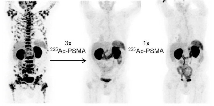
A revolutionary approach where the same radioactive molecule both finds cancer cells throughout your body and delivers a targeted dose of radiation to destroy them — sparing healthy tissue.
The word theranostics blends therapy and diagnostics. A specially designed molecule seeks out specific targets on cancer cells. First, a low-dose version is injected to image the tumour and confirm the target is present. Then, a therapeutic version delivers radiation directly to every site of disease — even spread invisible on conventional CT.
This precision means fewer side effects compared to traditional chemotherapy or external radiotherapy, and it can treat disease throughout the entire body in a single treatment cycle.
“We carry forward a legacy that began in 1957 when our founding pioneer, Dr. V.M. Sivaramakrishnan, established one of India’s first nuclear medicine centers. Today, we honor that vision by combining our six decades of experience with cutting-edge molecular imaging and therapeutic technologies. Our department stands at the unique intersection of physics, chemistry, and medicine—using the power of radiopharmaceuticals to see what others cannot see and treat what others cannot reach. Every nuclear medicine scan, every radionuclide therapy, and every diagnostic procedure we perform is driven by one commitment: to provide our patients with the most accurate diagnosis and effective treatment possible. We have a dedicated team of physicians, physicists, and technologists who work tirelessly to ensure that each patient receives personalized, evidence-based care with compassion and dignity.”
A small amount of radiotracer is injected. It binds to receptors on tumour cells. A PET/CT scan creates detailed 3D images showing every cancer deposit in the body.
The nuclear medicine team reviews your scan. If tumour uptake is confirmed, you are a candidate for therapy — the 'see before you treat' principle unique to theranostics.
The same molecule, now carrying a therapeutic isotope, is injected. It locks onto cancer cells and delivers a lethal radiation dose locally while decaying harmlessly elsewhere.
After each cycle, imaging is repeated to assess response. Treatment is adapted based on how the disease responds — a truly personalised approach.
Our department provides the full spectrum of radionuclide therapies, each precisely matched to the condition being treated.
When To Visit
Advanced dual-head gamma camera with SPECT capability for high-quality nuclear imaging of all organs
State-of-the-art nuclear medicine imaging system for precise diagnostic scans
Analyzes energy spectrum of gamma radiation for quality control and radiopharmaceutical purity testing
Instant Thin Layer Chromatography system for radiopharmaceutical quality assurance
High-quality medical image printing systems for producing diagnostic-grade hard copies of nuclear medicine scans
Intraoperative radiation detection device used during sentinel lymph node biopsy and tumor localization surgery
Dedicated isolation facility with proper radiation shielding for radioiodine therapy and other therapeutic nuclear medicine procedures
Comfortable patient rooms equipped with all necessary amenities and radiation safety features
Ventilated enclosure for safe handling of radioactive materials in patient care areas
Precisely measures radioactivity of radiopharmaceuticals before administration to ensure accurate patient dosing
Detects radioactive contamination on hands and feet of staff and patients for safety compliance
Personal radiation monitoring device worn by staff to track individual radiation exposure levels
Laminar flow workstations with LED lighting for safe handling and preparation of radiopharmaceuticals
Detects and measures radioactive contamination on surfaces and equipment
Continuously monitors ambient radiation levels in the department to ensure staff and patient safety
Shielded containers for safe disposal of radioactive waste materials
Personal protective equipment providing radiation shielding for staff during procedures
Technologist processing nuclear medicine images for diagnostic interpretation
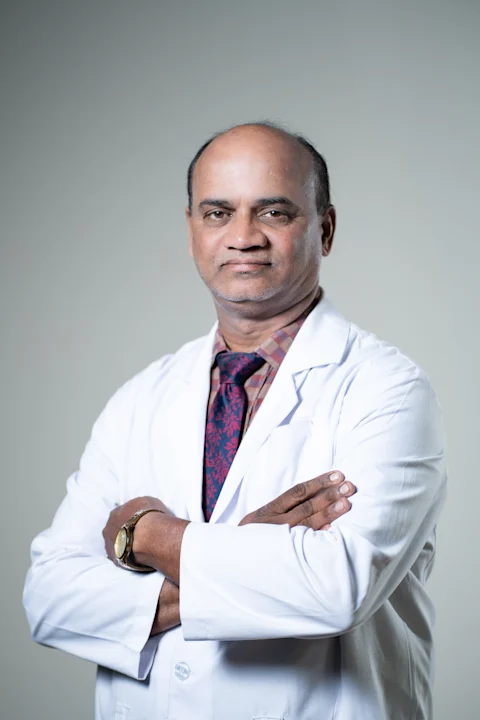
Consultant & Incharge - Department of Nuclear Medicine
Dr. Ashok Kumar R